X-rays
It is a form of electromagnetic radiation having a high frequency (energy) or shorter wavelength. It has no mass, no charge but has energy. It travels at the speed of light
When an X-ray is projected on the human body, three things happen i.e.
- Pass all the way through the body
- deflected or scattered
- be absorbed

X-rays passing through the tissues depends upon the energy and the atomic number of the tissue.
Higher energy X-ray pass through the body. On the other hand, higher atomic number things are more likely to absorb X-rays.
How does an X-ray create an image?
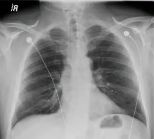
The X-ray that passes through the body to the film renders the film dark (Black). The X-ray that does not pass or blocked renders the film light (white).
Air = low atomic number = x-rays get through = image is dark (Black)
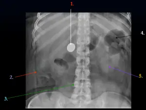
Metal = high atomic number = x-rays blocked = image is light (white)
X-ray pass through the body to varying degrees. This is due to the presence of different material inside the body such as air, fat, soft tissues, fluid, bones, metal, etc. These material act different to the action of the X-rays. This is defined in terms of Radiographic densities. The five basic Radiographic densities from black to bright white are given below.
- Air
- Fat
- Soft tissues/fluid
- Mineral/Bones
- Metal

The X-rays completely passes through the Air that renders the film, Black. and completely absorb the metal that renders the film, White. From top to bottom of the above list, film colour changes from Black to White.
Medical Imaging
- The primary purpose is to identify pathologic conditions.
- Requires recognition of normal anatomy.
Radiographic Analysis
Any structure, normal or pathologic should be analysed for
- Size
- Shape & Colour
- Position
- Density (5 basic radiographic densities as discussed above)
Excercise
Q1. How do x-rays create an image of internal body structures?
Ans.
- X-rays pass through the body to varying degrees
- Higher atomic number structures block x-rays better, example bone.
- Lower atomic number structures allow x-rays to pass through, example: air in the lungs.
Q2. What are the 5 basic radiographic densities?
Ans. The five basic Radiographic densities are given below.
- Air
- Fat
- Soft tissues/fluid
- Mineral/Bones
- Metal
Q3. What three things can you do to protect yourself from radiation?

I have exam today and this saves me. Please add more points and questions to this article.
Good for students having bio-instrumentation course. Short and to the point notes.
these notes are good for quick revision. Thanks alot.