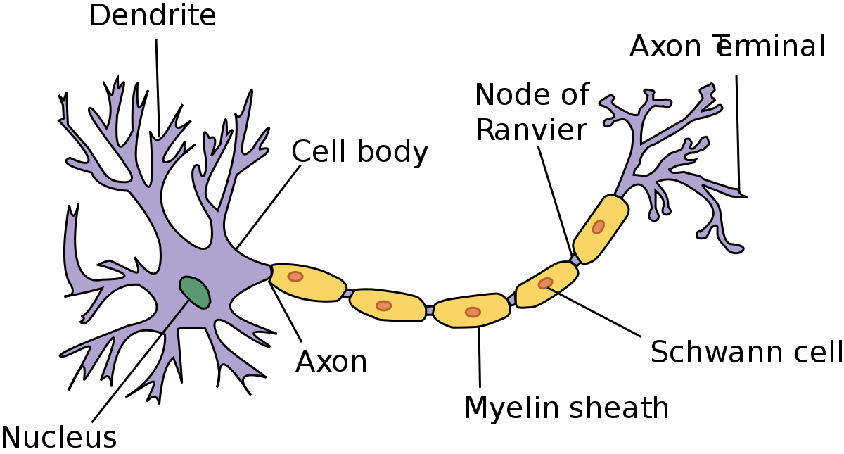
The long tail like structure connected with the cell body is known as Axon. The portion of the axon immediately adjacent to the cell body is called the axon hillock. Branches like structure connected to the cell body are known as Dendrites.
Axons and dendrites are coated with a fatty insulating substance called Myelin. This coating is known as Myelin sheath.
Myelin sheath is interrupted at regular intervals by Nodes of Ranvier. It helps in speed transmission of information along the nerves.
Outside nervous system, the myelin sheath is covered with another insulating layer, called Neurilemma. It is made up of thin cells, called Schwann cells.
The main difference between axon and dendrites is the function of the fiber and the direction in which it carries information with respect to the cell body.
Both axons and dendrites are called as Nerve fibers.
A nerve is the bundle of individual nerve fibers.
Afferent Nerves: Nerves that Carry sensory information from the various parts of the body to the brain.
Efferent Nerves: Nerves that carry signals from the brain to operate various muscles.
Central Nervous System (CNS)
It consists of the brain and the spinal cord. CNS contains a large number of neurons of variety and large fatty cell bodies called Glial cells.
Glial cells play an important role in ridding the brain of foreign substances and memory function.
Gray matter = cell bodies plus small fibers in brain = Grey in colour
White matter = Myelin coated large fibers = White in colour
The collection of neuronal cell bodies in CNS is known as Nuclei. Similar collection outside CNS is known as Ganglia. CNS is bilaterally symmetrical. Several functions of CNS are crossed over. Left side brain control right side body. Right side brain control left side body.Peripheral Nerves: Nerve fibers outside the CNS.
Pheripheral Nerves = Afferent + Efferent nerves
Sensory Nerves: Afferent Peripheral Nerves that bring sensory information into CNS.
Motor Nerves: Efferent Peripheral Nerves that control the motor functions of muscles.
The interconnections between neurons are called synapses. All synapses occur at or near cell bodies. Synapse don’t touch each other but do come into close proximity so that the axon (output) of one nerve can activate the dendrites or cell body (input) of another by producing a chemical that stimulates the membrane of a dendrite or cell body.
Note: The communication can take place in one direction only in neurons.
The block diagram of the nervous system is shown below. CNS and peripheral nervous system are part
Fig. Nervous System block diagram
Image Source: classconnection.s3.amazonaws.com
Parts of the Brain
image source: askabiologist.asu.edu
Function of parts of the Brain
1. Medulla
Controls basic function responsible for life, such as breathing, heart rate, and kidney function.
2. Pons
It is an interconnecting area. It contains nuclei which play an important role in salvation, feeding and facial expression. It contains relay for the auditory system, spinal motor neurons, and some respiratory nuclei.
3. Cerebellum
Physiological microcomputer which intercepts various sensory and motor nerves to smooth out muscle motion. The cerebellum also plays a vital role in man’s ability to maintain his balance.
4. Thalamus
Manipulate all sensory information on its way to the cerebrum.
5. Reticular activation system (RAS)
It keeps a person awake and alert and causes him/her to pay attention to a sensory input.
6. Hypothalamus
It is the center for emotions in the brain. It controls the neural regulation of endocrine gland functions via the pituitary gland and contains nuclei responsible for eating, drinking, etc.
The brain can be divided into two halves called the cerebral hemispheres. Each cerebral hemisphere is divided into four lobes by sulci and gyri.
The sulci or fissures are the grooves and the gyri are the bumps that can be seen on the surface of the brain.
Cerebrum is divided into 4 sectors i.e.
1. Frontal lobe
Located in front of the central sulcus. Associated with reasoning, planning, parts of speech, movement, emotions, and problem-solving.
2. Parietal lobe
Located behind the central sulcus. Associated with movement, orientation, recognition, perception of stimuli
3. Occipital lobe
Located at the back of the brain, behind the parietal lobe and temporal lobe. Associated with visual processing.
4. Temporal lobe
Located below the lateral fissure. Associated with perception and recognition of auditory stimuli, memory, and speech.